A revolution in Radiology
The digitizing of X-ray imaging will improve the images. However, digital post-processing will depend on the quality of original the X-ray image. MIQvet calculates the optimal settings for kV and mAs of your X-ray device when making images. With MIQvet images will show the details needed for making the diagnosis.
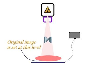
The original image
The original image is set as soon as radiation leaves the patient. In film imaging this is the final image since postprocessing doesn't exist. The image on film cannot be edited.
In digital imaging editing is possible. With the aid of a computer different levels of detected radiation in the original image can be emphasized and thus interpretation will be easier. The range of shades can be set from white to black.
Manipulating the image is called postprocessing.

Post-processing has limitations
In digital imaging the computer offers an option in the editing of X-ray image. However, a computer can only emphasize differences in gray scales which are already present in the original image. When there is no contrast in the original image, the computer cannot add it: all pixels in the same shade of gray will be manipulated in the same way.
In addition, software can only reduce the number of shades by merging them and thus reduce contrast. This will result in a loss of details.
MIQvet adjusts
MIQvet will create optimal circumstances for the original image. By optimizing the settings for kV and mAs the minimum dose of radiation and the optimal kV setting will be guaranteed.
As a result postprocessing will be more effective due to more contrast in the original image and details will be more evident. This will facilitate interpretation and lead to a more accurate diagnosis.
MIQvet makes the difference
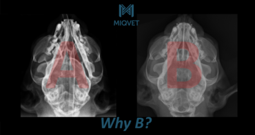
Watch these images. Everything was the same except settings for kV and mAs.
Both X-ray images range from completely white to completely black. This is done by the computer. The image on the left obviously has less shades of gray than the right image. In extremis you would say it's either white or black, either yes or no. The image on the right is more grayish and will show more details.
The number of different colors in the image on the left is less than in the image on the right as a result the image will be more black and white and less gray. The difference from one color to the next one is greater by the merging of colors. Result is a loss of details and the image will look grainish.

The details
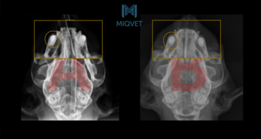
The image on the right side has more different shades of gray. There is more contrast and there are more nuances in bone structure. The soft tissues are better displayed which is evident in the nostrils. In the left image the conchae are not visible, in the right image they can easily be distinguished.

Diversity in grayscales is the solution
Since on the left image the colors in the bone tissue are very close together, few distinctions can be made between sections. The editing program can not solve this lack of contrast. Software can only emphasize the existing contrast in the image. An image with many shades of gray is therefore the solution for better imaging.
What happens when colors are merged can be seen in this image. With 20 scales a decent image can be made by the computer. When only 3 scales are available there will be an enormous gap between adjacent scales. In the end with 2 scales, pixels will be either black or white, this is called the pencil drawing.

Finding the right settings is a sensitive balance
By correctly setting mAs and kV of the X-ray tube depending on your subject, there will be more different colors of gray in the image. The computer can emphasize the contrast by manipulating adjacent pixels with different values for their gray scalesand and details will be visable.
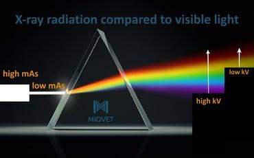
It is similar to photography: there are 2 options to manipulate the image with the camera. With the opening of the diaphragm the amount of light is regulated and the color of the light is daylight or flashlight.
In X-ray imaging, mAs is the parameter for the "amount of light" and kV is the parameter for the "color of light".
The issue in digital radiology is that underexposure (by definition due to too not enough mAs) and overexposure (by definition due to too much kV) both produce identical, grainy and unbalanced images. The question is therefore: what to look at? Is it the mAs or the kV? MIQvet solves this problem and determines the optimum balance between patient, mAs and kV.
Learn more about MIQvet


